Autonomous Sensory Zones
(peripheral nerves)
Median nerve illustration by Guttmann (1939)
3) Sensory zones
a.maximal zone: maximal area supplied by a peripheral nerve
- maximal=intermediate+autonomous
b.intermediate zone: area of overlap of the maximal zone of different peripheral nerves
c.autonomous zone: area exclusively supplied by a particular peripheral nerve
4) Autonomous zones of various nerves
a. Radial nerve
b. Median nerve
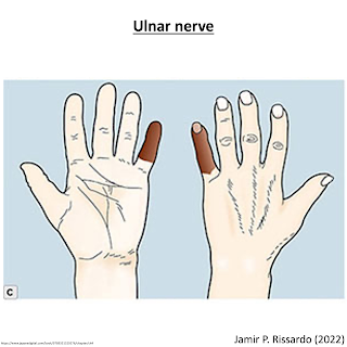
c. Ulnar nerve
d. Common peroneal nerve
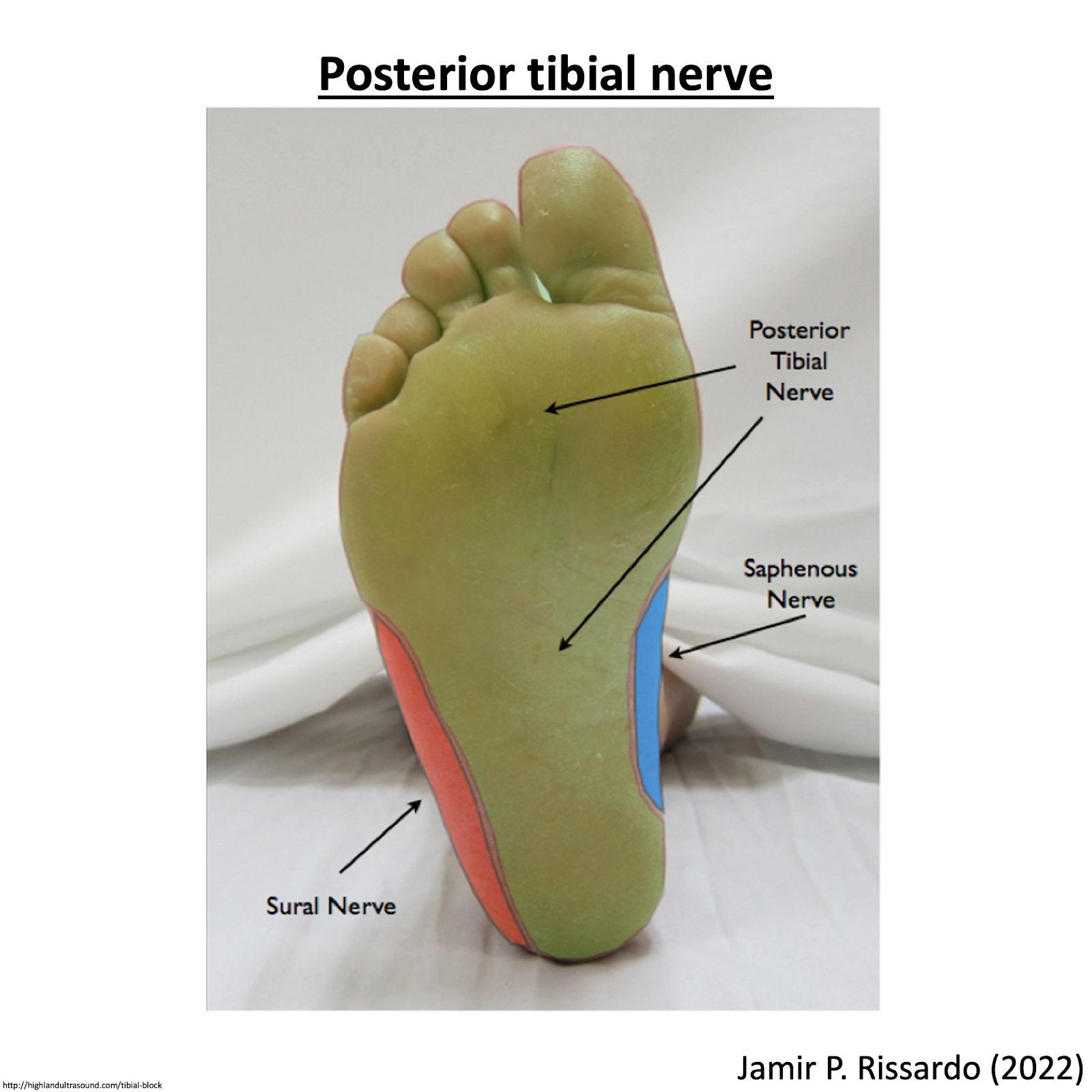
e. Sciatic nerve
5) Radial nerve
area: 1st dorsal web space of hand (Anatomical snuff box)
“according to some authors, radial nerve and common peroneal do not have autonomous zones although complete transection of the nerve results in sensory loss over the mentioned regions”
7) Ulnar nerve
area: distal phalanx (tip) of little finger (5th finger)
8) Common peroneal nerve
area: central strip on dorsum of foot
“according to some authors, radial nerve & common peroneal do not have autonomous zones although complete transection of the nerve results in sensory loss over the mentioned regions”
13) MRC grading


