Article type: Literature Review
Article title: Cardiac 123I-Metaiodobenzylguanidine (MIBG) Scintigraphy in Parkinson’s Disease: A Comprehensive Review
Journal: Brain Sciences
Year: 2023
Authors: Jamir Pitton Rissardo and Ana Letícia Fornari Caprara
E-mail: jamirrissardo@gmail.com
ABSTRACT
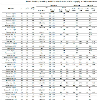
Cardiac sympathetic denervation, as documented on 123I-metaiodobenzylguanidine (MIBG) myocardial scintigraphy, is relatively sensitive and specific for distinguishing Parkinson’s disease (PD) from other neurodegenerative causes of parkinsonism. The present study aims to comprehensively review the literature regarding the use of cardiac MIBG in PD. MIBG is an analog to norepinephrine. They share the same uptake, storage, and release mechanisms. An abnormal result in the cardiac MIBG uptake in individuals with parkinsonism can be an additional criterion for diagnosing PD. However, a normal result of cardiac MIBG in individuals with suspicious parkinsonian syndrome does not exclude the diagnosis of PD. The findings of cardiac MIBG studies contributed to elucidating the pathophysiology of PD. We investigated the sensitivity and specificity of cardiac MIBG scintigraphy in PD. A total of 54 studies with 3114 individuals diagnosed with PD were included. The data were described as means with a Hoehn and Yahr stage of 2.5 and early and delayed registration H/M ratios of 1.70 and 1.51, respectively. The mean cutoff for the early and delayed phases were 1.89 and 1.86. The sensitivity for the early and delayed phases was 0.81 and 0.83, respectively. The specificity for the early and delayed phases were 0.86 and 0.80, respectively.
Keywords: neuroimaging; cardiac imaging; myocardium; 123I; 131I; pre-motor dysfunction; non-motor dysfunction; autonomic dysfunction; parkinsonism; movement disorder
Full text available at:
DOI
10.3390/brainsci13101471
Citation
Rissardo JP, Caprara ALF. Cardiac 123I-Metaiodobenzylguanidine (MIBG) Scintigraphy in Parkinson's Disease: A Comprehensive Review. Brain Sci 2023;13:1471.
Figure 1. The two types of metaiodobenzylguanidine (MIGB) uptake into cells. Type 1 is an active pathway. Type 2 is a passive pathway by simple diffusion.
Figure 2. Cardiac MIBG results in different neurological conditions. Abbreviations: DLB, dementia with Lewy bodies; H/M, heart-to-mediastinum ratio; PAF, pure autonomic failure; PD, Parkinson’s disease; RBD, rapid eye movement sleep behavior disorder.
Figure 3. Polar map of cardiac perfusion. (A) Vertical long axis; (B) short axis; and (C) left ventricular (LV) segmentation. Coronary artery territories: left anterior descending artery (dark green); right coronary artery (gray); left circumflex artery (white). Segments: (1) basal anterior; (2) basal anteroseptal; (3) basal inferoseptal; (4) basal inferior; (5) basal inferolateral; (6) basal anterolateral; (7) mid anterior; (8) mid anteroseptal; (9) mid inferoseptal; (10) mid inferior; (11) mid inferolateral; (12) mid anterolateral; (13) apical anterior; (14) apical septal; (15) apical inferior; (16) apical lateral; (17) apex.
Table 1. Interference from some drugs in the uptake of MIBG by Solanki et al. [20] modified by Rissardo et al.
Table 2. Sensitivity, specificity, and H/M ratio of cardiac MIBG scintigraphy in Parkinson’s disease.


