Article type: Case Report and Literature Review
Article title: Herpes zoster ophthalmicus presenting with orbital myositis prior to the appearance of vesicular lesions: A case report and mini‑review of the literature
Journal: Geriatrics
Year: 2024
Authors: Jamir Pitton Rissardo, Pranav Patel, and Ana Letícia Fornari Caprara
E-mail: jamirrissardo@gmail.com
ABSTRACT
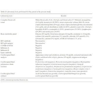
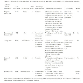
All orbital tissues, including extra-ocular muscles, can be affected by the varicella-zoster virus (VZV). However, only a minority of all individuals with herpes zoster infections present with herpes zoster ophthalmicus. The present study reports the case of a middle-aged male patient presenting with an acute intractable right-sided headache. His neurological examination yielded normal results. The analysis of cere-brospinal fluid by biochemistry and cultural analysis yielded normal results; however, the analysis of this fluid using poly-merase chain reaction yielded a positive result for VZV. Thus, treatment with acyclovir was commenced. Brain magnetic resonance imaging revealed a bilateral intraorbital intraconal enhancement consistent with myositis. His symptoms evolved into a shock-like pain over the scalp associated with painful ocular movements. On the 2nd day of admission, he developed new vesicular lesions found on the right-side cranial nerve V1 dermatome. By the 6th day of admission, he was asymptom-atic, and his physical examination revealed the resolution of the dermatologic manifestations of the VZV. The patient was stable for outpatient follow-up with ophthalmology and was discharged on an oral valacyclovir course for 7 days. To the authors' knowledge, there are four cases reported in the literature of herpes zoster ophthalmicus with orbital myositis prior to the appearance of vesicular lesions. Thus, it is suggested that VZV serology be investigated before a final diagnosis of idiopathic orbital myositis is made.
Keywords: Key words: herpes zoster, varicella zoster, varicella‑zoster virus, orbital syndrome, orbital myositis, herpes zoster ophthalmicus, shingles, immunocompetent
Full text available at:
DOI
Citation
Pitton Rissardo J, Patel P, Fornari Caprara AL. Herpes zoster ophthalmicus presenting with orbital myositis prior to the appearance of vesicular lesions: A case report and mini‑review of the literature. Med Int 2024;4:(6)61.
Figure 1. Brain magnetic resonance imaging illustrating myositis of the right extraocular muscles. Patchy bilateral intraorbital intraconal enhancement around the optic nerve sheath complexes and asymmetric thickening and enhancement of the right extraocular muscles. (A) Axial T1‑weighted, (B) fluid attenuated inversion recovery, (C) T2‑weighted fat suppressed, and (F) T1‑weighted turbo spin echo images; (D) coronal T2‑weighted and (E) T1‑weighted turbo spin echo images are shown.
Figure 2. Anatomical view of the forehead showing vesicular lesions on the 2nd day of admission. The lesions crusted within 2 weeks at the follow‑up and disappeared within 1 month.
Table I. Laboratory tests performed for the patient in the present study.
Table II. Cases reported in the literature of orbital myositis preceding skin symptoms in patients with varicella zoster infection.


